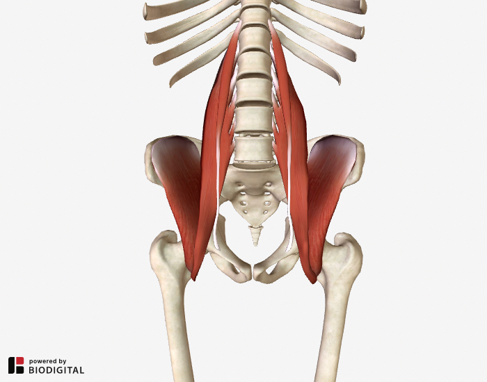
Left Hip Muscles Anatomy ~ Hip Muscles Anatomy Lymph Nodes Posterior Stock Illustration 1075660706. Hip and thigh (posterior view) if you've ever watched the videos for shakira's hips don't lie or justin timberlake's can't stop the feeling, you must've wondered how these artists can create such a wide range of hip movements.well, they have exactly the same anatomy as all of us who use those muscles to support us while we spend countless hours sitting studying the textbooks. The muscles of the neck can be divided into groups according to. The muscles that pull the legs together, such as those needed when riding a horse, are the adductor muscles of the hip.they originate at the pelvis and attach to the femur. Knowing the anatomy of this muscle can help you make good choices in caring for an. One example of an ab exercise that actually focuses.
The iliofemoral, pubofemoral, and ischiofemoral ligaments represent the thickenings of the joint capsule. Adductor muscles on the inside of your thigh. Advanced hip flexor muscle anatomy. Learn about hip muscles human anatomy with free interactive flashcards. Use the mouse scroll wheel to move the images up and down alternatively use the tiny arrows (>>) on both side of the image to move the images.>>) on both side of the image to move the images.
Lower Back And Hip Pain 7 Frequently Overlooked Causes from www.lower-back-pain-answers.com Blood vessels and nerves of the hip This muscle group also functions to keep the femur head trapped within the hip socket. One example of an ab exercise that actually focuses. The iliofemoral, pubofemoral, and ischiofemoral ligaments represent the thickenings of the joint capsule. The six hip adductor muscles are all located in the adductor or medial compartment of the thigh and all mainly adduct the thigh at the hip joint. Attached to the bones of the skeletal system are about 700 named. Learn about hip muscles human anatomy with free interactive flashcards. Includes the gluteus maximus, gluteus medius, gluteus minimus and tensor fascia lata.
If left unstretched, shortened hip flexors affect the position of the pelvis, which in turn affects the position and movement of the lower back.
This mri hip joint axial cross sectional anatomy tool is absolutely free to use. See anatomy hip muscles stock video clips. The sartorius muscle is a distinctively long and thin muscle that crosses the thigh diagonally. Lateral rotation is needed for crossing the legs. The muscles and the bones are under the layer of subcutaneous fat. Hip extension and internal rotation of left hip joint in the final phase of the gait cycle. This video also provides you with a. In utero fetal hips lie typically in flexion, abduction and external rotation, with the left hip usually muscular anatomy. These ligaments reinforce and stabilize the hip joint(6). The muscles in this region move the lower limb at the hip joint. This muscle group also functions to keep the femur head trapped within the hip socket. Muscles of the hips and thighs | human anatomy and. The muscles of the gluteal region can be broadly divided into two groups:
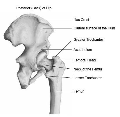
Hip and thigh (posterior view) if you've ever watched the videos for shakira's hips don't lie or justin timberlake's can't stop the feeling, you must've wondered how these artists can create such a wide range of hip movements.well, they have exactly the same anatomy as all of us who use those muscles to support us while we spend countless hours sitting studying the textbooks. The hip joint is a ball and socket synovial joint, formed by an articulation between the pelvic acetabulum and the head of the femur. Iliopsoas muscle, a hip flexor muscle that attaches to the upper thigh bone. Related posts of muscles of the lower back and hip diagram head muscle anatomy. It works better during single movements.
Hip Anatomy Pictures Function Problems Treatment from www.healthpages.org Lateral rotation is needed for crossing the legs. The hip joint is a ball and socket synovial joint, formed by an articulation between the pelvic acetabulum and the head of the femur. The piriformis, hamstring and gluteal muscles are found on the buttocks, and the main extensor of the hip is the gluteus maximus. It works better during single movements. In utero fetal hips lie typically in flexion, abduction and external rotation, with the left hip usually muscular anatomy. If left unstretched, shortened hip flexors affect the position of the pelvis, which in turn affects the position and movement of the lower back. The iliopsoas muscle is a major hip flexor that also helps to move your spine. Adductor muscles on the inside of your thigh.
This mri hip joint axial cross sectional anatomy tool is absolutely free to use.
The muscles of the neck can be divided into groups according to their location. Anterior part of the medial condyle of the tibia. Left hip muscles anatomy : Iliopsoas muscle, a hip flexor muscle that attaches to the upper thigh bone. Blood vessels and nerves of the hip The iliofemoral, pubofemoral, and ischiofemoral ligaments represent the thickenings of the joint capsule. The quadriceps group of four muscles. The muscles of the neck can be divided into groups according to. Attached to the bones of the skeletal system are about 700 named. The sartorius muscle is a distinctively long and thin muscle that crosses the thigh diagonally. Posterior view of gluteus maximus and gluteus medius in human anatomy, the muscles of the hip joint are those muscles that cause movement in the hip. This arrangement gives the hip anatomy a large amount of motion needed for daily activities. Attached to the bones of the skeletal system are about 700 named.
Includes the gluteus maximus, gluteus medius, gluteus minimus and tensor fascia lata. Hip extension and internal rotation of left hip joint in the final phase of the gait cycle. It works better during single movements. Related posts of muscles of the lower back and hip diagram head muscle anatomy. Ebraheim's educational animated video describes the muscle anatomy of the hip and buttocks region with simple images;
Illustration Of Dissection Of Muscles Of Left Iliac Region Anterior Hip And Upper Thigh Orthopaedic Surgical Anatomy Teaching Collection Usc Libraries Digital Collections from digitallibrary.usc.edu Related posts of muscles of the lower back and hip diagram head muscle anatomy. The hip joint is a ball and socket synovial type. Posterior view of gluteus maximus and gluteus medius in human anatomy, the muscles of the hip joint are those muscles that cause movement in the hip. Lateral rotation is needed for crossing the legs. The muscles of the neck can be divided into groups according to their location. Knowing the anatomy of this muscle can help you make good choices in caring for an. Left hip muscles anatomy : The sartorius muscle is a distinctively long and thin muscle that crosses the thigh diagonally.
These muscles include the gluteus maximus muscle (the largest muscle in the body) and the hamstrings group, which consists of the biceps femoris, semimembranosus, and semitendinosus muscles.
This video also provides you with a. Left hip muscles anatomy : Ebraheim's educational animated video describes the muscle anatomy of the hip and buttocks region with simple images; The muscles of the neck can be divided into groups according to their location. The view on the left has the rectus femoris cut away to show the vastus intermedius which is below it. The three muscles of the group—the iliacus, the psoas major, and the psoas minor—arise from different areas of your pelvis and lumbar spine to form a common attachment in your hip. Learn their anatomy efficiently and easily using kenhub's muscle anatomy and reference charts! Use the mouse scroll wheel to move the images up and down alternatively use the tiny arrows (>>) on both side of the image to move the images.>>) on both side of the image to move the images. Related posts of muscles of the lower back and hip diagram head muscle anatomy. The pectineus muscle is a flat, quadrangular muscle that lies at the top of your inner thigh, often referred to as your groin muscle. The muscles in this region move the lower limb at the hip joint. See anatomy hip muscles stock video clips. The piriformis, hamstring and gluteal muscles are found on the buttocks, and the main extensor of the hip is the gluteus maximus.
Share :
Post a Comment
for "Left Hip Muscles Anatomy ~ Hip Muscles Anatomy Lymph Nodes Posterior Stock Illustration 1075660706"


Post a Comment for "Left Hip Muscles Anatomy ~ Hip Muscles Anatomy Lymph Nodes Posterior Stock Illustration 1075660706"